What are the three types of salivary glands and where are they located in the mouth?
A. Parotid, sublingual, and submandibular glands located in the cheeks, tongue, and roof of the mouth, respectively.
B. Sublingual, submandibular, and buccal glands located in the tongue, cheeks, and lips, respectively.
C. Parotid, sublingual, and submandibular glands located in the roof of the mouth, cheeks, and under the jawbone, respectively.
D. Sublingual, parotid, and buccal glands located in the tongue, cheeks, and lips, respectively.
The three major pairs of salivary glands are the parotid glands, sublingual glands, and submandibular glands.
- Parotid glands are located just in front of your ears.
- Sublingual glands are located below either side of your tongue, under the floor of your mouth.
- Submandibular glands are located below your jaw.
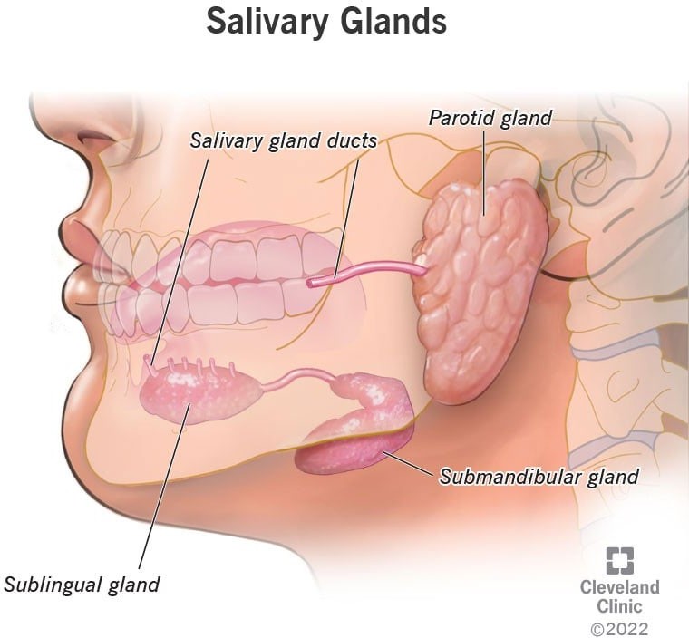 |
Therefore, the Correct Answer is C.
More Questions on TEAS 7 Science
-
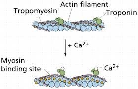
Q #1: What is the role of calcium in muscle contraction?
A. Calcium binds to tropomyosin to expose the myosin-binding sites on actin.
B. Calcium is released from the sarcoplasmic reticulum to initiate the sliding of actin and myosin filaments.
C. Calcium activates the motor neurons to stimulate muscle contraction.
D. Calcium is required for the relaxation of muscles after contraction.
Answer Explanation
Muscle contraction is a complex process that involves the interaction between actin and myosin filaments in the muscle fibers. The sliding of these filaments is initiated by the release of calcium ions from the sarcoplasmic reticulum, a specialized organelle in muscle cells. The calcium ions bind to the protein troponin, which causes a conformational change in the troponin-tropomyosin complex, exposing the myosin-binding sites on actin. This allows the myosin heads to bind to actin, forming cross-bridges that pull the actin filaments towards the center of the sarcomere, resulting in muscle contraction.
Option a) is incorrect because calcium does not bind to tropomyosin directly, but rather binds to the protein troponin, causing a conformational change in the troponin-tropomyosin complex. Option c) is incorrect because calcium does not activate motor neurons, but rather is released from the sarcoplasmic reticulum in response to an action potential that travels down the motor neuron to the neuromuscular junction. Option d) is incorrect because calcium is required for muscle contraction, not relaxation. The relaxation of muscles after contraction is due to the active transport of calcium ions back into the sarcoplasmic reticulum, which allows the troponin-tropomyosin complex to return to its resting conformation, blocking the myosin-binding sites on actin and ending the cross-bridge cycle.
-
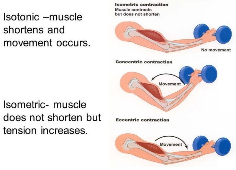
Q #2: What is the difference between isotonic and isometric muscle contractions?
A. Isotonic contractions produce no movement while isometric contractions produce movement.
B. Isotonic contractions produce movement while isometric contractions produce no movement.
C. Isotonic contractions generate tension in the muscle while isometric contractions involve shortening of the muscle fibers.
D. Isotonic contractions involve contraction of individual muscle fibers while isometric contractions involve the entire muscle.
Answer Explanation
Isotonic and isometric contractions are two types of muscle contractions that differ in the amount of force produced and the movement of the muscle. In isotonic contractions, the muscle changes length and produces movement, such as lifting a weight. The force generated by the muscle remains constant throughout the movement. Isotonic contractions can be further classified as concentric contractions, in which the muscle shortens as it contracts, and eccentric contractions, in which the muscle lengthens as it contracts.
In contrast, isometric contractions occur when the muscle generates force without changing its length or producing movement. For example, holding a weight in a fixed position without moving it requires an isometric contraction. In an isometric contraction, the force generated by the muscle increases up to a maximum and then remains constant. Isometric contractions can be used to build strength and endurance in the muscle, but they do not produce movement.
-
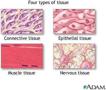
Q #3: Which of the following is NOT one of the four primary tissue types found in the human body?
A. Epithelial
B. Nervous
C. Connective
D. Exocrine glandular
Answer Explanation
Exocrine glandular is not one of the four primary tissue types found in the human body. The four primary tissue types are epithelial, nervous, connective, and muscle.
-
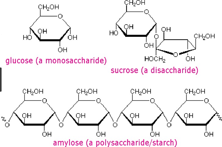
Q #4: What is the difference between a monosaccharide and a disaccharide?
A. Monosaccharides are composed of two sugar molecules while disaccharides are composed of a single sugar molecule.
B. Monosaccharides are simple sugars that cannot be further broken down into simpler sugars while disaccharides are composed of two simple sugars.
C. Monosaccharides are only found in plants while disaccharides are only found in animals.
D. Monosaccharides are used for energy storage while disaccharides are used for structural purposes.
Answer Explanation
Carbohydrates are one of the main types of biomolecules and are composed of monomers called monosaccharides. Monosaccharides are simple sugars that cannot be further broken down into simpler sugars. They are usually composed of 3 to 7 carbon atoms and have a general formula of (CH2O)n, where n is a number between 3 and 7. Examples of monosaccharides include glucose, fructose, and galactose.
When two monosaccharides are joined together by a glycosidic bond, they form a disaccharide. Disaccharides are composed of two simple sugars and can be broken down into their constituent monosaccharides by hydrolysis. Examples of disaccharides include sucrose, lactose, and maltose.
Option a) is incorrect because it describes the composition of a disaccharide, not a monosaccharide. Option
c) is incorrect because both monosaccharides and disaccharides can be found in both plants and animals.
Option d) is incorrect because both monosaccharides and disaccharides can be used for energy storage and
structural purposes, depending on their specific structure and function in the organism.
-
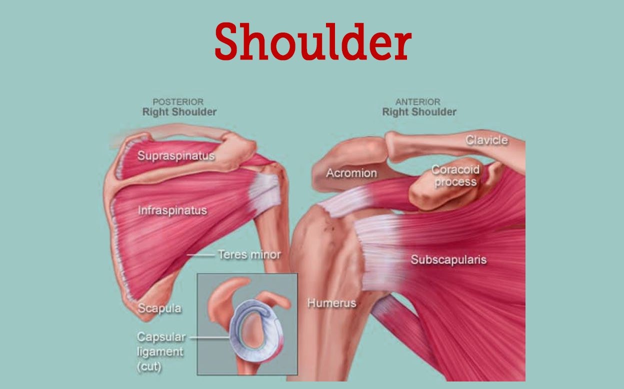
Q #5: What is the name of the joint that allows for rotation of the arm at the shoulder?
A. Elbow joint
B. Hip joint
C. Knee joint
D. Shoulder joint
Answer Explanation
-
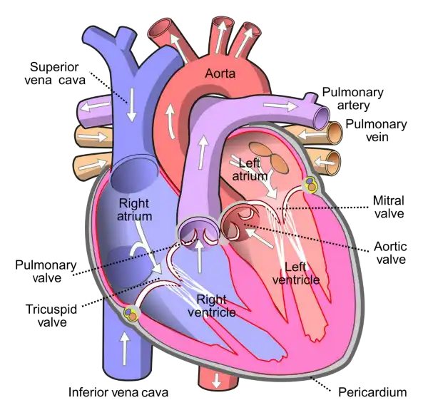
Q #6: What is the name of the valve that separates the left atrium and left ventricle in the heart?
A. Aortic valve
B. Mitral valve
C. Tricuspid valve
D. Pulmonary valve
Answer Explanation
The mitral valve is located between the left atrium and left ventricle of the heart and helps to regulate the flow of blood between these chambers. It consists of two leaflets or flaps that open and close in response to changes in pressure as the heart beats.
During diastole, when the heart is relaxed and filling with blood, the mitral valve opens to allow blood to flow from the left atrium into the left ventricle. During systole, when the heart contracts to pump blood out of the left ventricle and into the systemic circulation, the mitral valve closes to prevent backflow of blood into the left atrium.
The mitral valve is one of four valves in the heart that help to ensure the unidirectional flow of blood through the heart and the rest of the circulatory system. Problems with the mitral valve, such as mitral valve prolapse or mitral stenosis, can lead to a range of symptoms and complications, including shortness of breath, fatigue, chest pain, and heart failure.
-
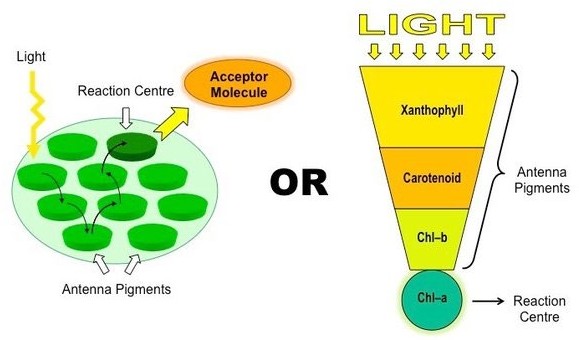
Q #7: What is the primary pigment responsible for photosynthesis in plants?
A. Chlorophyll a
B. Chlorophyll b
C. Carotenoids
D. Anthocyanins
Answer Explanation
Chlorophyll a is the primary pigment responsible for photosynthesis in plants. It is a green pigment that is essential for capturing light energy from the sun and converting it into chemical energy that can be used by the plant. Chlorophyll a absorbs light most efficiently in the blue and red parts of the spectrum, and reflects green light, giving plants their characteristic green color
Chlorophyll b is another type of chlorophyll that is also involved in photosynthesis, but it is not as abundant as chlorophyll a. Chlorophyll b absorbs light most efficiently in the blue and orange parts of the spectrum and reflects yellow-green light.
Carotenoids are pigments that are present in many plants and are involved in photosynthesis as well as protecting the plant from damage caused by excess light. Carotenoids are responsible for the orange, yellow, and red colors of many fruits and vegetables.
Anthocyanins are pigments that give plants their red, purple, and blue colors. While they are not directly involved in photosynthesis, they play a role in atracting pollinators and protecting the plant from damage caused by UV radiation.
-
Q #8: What is the difference between innate immunity and adaptive immunity?
A. Innate immunity is present at birth and provides immediate, non-specific protection against pathogens while adaptive immunity is developed over time and provides specific protection against particular pathogens.
B. Innate immunity involves the recognition of specific pathogens while adaptive immunity involves the recognition of general paterns of pathogens.
C. Innate immunity involves the production of antibodies while adaptive immunity involves the activation of phagocytes.
D. Innate immunity is activated by the lymphatic system while adaptive immunity is activated by the circulatory system.
Answer Explanation
Innate immunity is the first line of defense against pathogens and is present at birth. It provides immediate, non-specific protection against a wide range of pathogens, including bacteria, viruses, and fungi. Innate immunity involves physical barriers, such as skin and mucous membranes, as well as cellular and molecular components, such as phagocytes and cytokines.
Adaptive immunity, on the other hand, is developed over time and provides specific protection against particular pathogens. It involves the recognition of antigens, which are specific components of pathogens, by immune cells called lymphocytes. The lymphocytes then produce antibodies that are specific to the antigens, allowing for a targeted response to the pathogen. This process takes time to develop, as the immune system needs to encounter the pathogen and mount a response.
Overall, innate immunity provides immediate, non-specific protection while adaptive immunity provides specific protection that is tailored to the particular pathogen. Both forms of immunity work together to protect the body against pathogens.
-
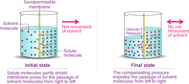
Q #9: Which of the following describes the process of osmosis?
A. Movement of substances from an area of high concentration to an area of low concentration.
B. Movement of substances against a concentration gradient with the help of transport proteins.
C. Movement of water molecules from an area of high concentration to an area of low concentration through a selectively permeable membrane.
D. Movement of substances into a cell by engulfing them with the plasma membrane.
Answer Explanation
Osmosis is the process by which water molecules move across a selectively permeable membrane from an area of high concentration to an area of low concentration, in order to equalize the concentration of solutes on both sides of the membrane. Selectively permeable membranes allow only certain molecules to pass through, while preventing the passage of others.
In osmosis, the movement of water molecules is driven by the concentration gradient of solutes, which cannot pass through the membrane. If one side of the membrane has a higher concentration of solutes than the other, water molecules will move from the side with the lower concentration of solutes to the side with the higher concentration of solutes, in an atempt to dilute the solutes and equalize the concentration on both sides.
Osmosis is important in many biological processes, including the uptake of water by plant roots, the regulation of water balance in animal cells, and the preservation of food by adding salt or sugar to create a hypertonic environment that inhibits bacterial growth.
-
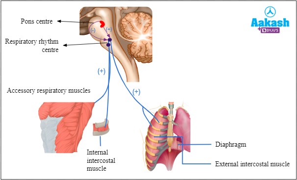
Q #10: Which part of the respiratory system is responsible for regulating breathing rate and depth?
A. Bronchi
B. Alveoli
C. Diaphragm
D. Trachea
Answer Explanation
Diaphragm is responsible for regulating breathing rate and depth. It is a dome-shaped muscle located at the
bottom of the chest cavity that contracts and relaxes to help move air in and out of the lungs.