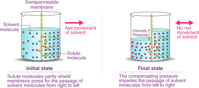
Which of the following describes the process of osmosis?
A. Movement of substances from an area of high concentration to an area of low concentration.
B. Movement of substances against a concentration gradient with the help of transport proteins.
C. Movement of water molecules from an area of high concentration to an area of low concentration through a selectively permeable membrane.
D. Movement of substances into a cell by engulfing them with the plasma membrane.
Osmosis is the process by which water molecules move across a selectively permeable membrane from an area of high concentration to an area of low concentration, in order to equalize the concentration of solutes on both sides of the membrane. Selectively permeable membranes allow only certain molecules to pass through, while preventing the passage of others.
In osmosis, the movement of water molecules is driven by the concentration gradient of solutes, which cannot pass through the membrane. If one side of the membrane has a higher concentration of solutes than the other, water molecules will move from the side with the lower concentration of solutes to the side with the higher concentration of solutes, in an atempt to dilute the solutes and equalize the concentration on both sides.
Osmosis is important in many biological processes, including the uptake of water by plant roots, the regulation of water balance in animal cells, and the preservation of food by adding salt or sugar to create a hypertonic environment that inhibits bacterial growth.
 |
Therefore, the Correct Answer is C.
More Questions on TEAS 7 Science
-
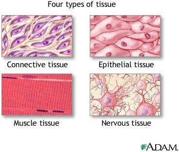
Q #1: Which of the following is NOT one of the four primary tissue types found in the human body?
A. Epithelial
B. Nervous
C. Connective
D. Exocrine glandular
Answer Explanation
Exocrine glandular is not one of the four primary tissue types found in the human body. The four primary tissue types are epithelial, nervous, connective, and muscle.
-
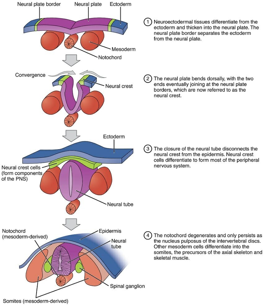
Q #2: During embryonic development, which of the following germ layers forms the nervous system?
A. Ectoderm
B. Endoderm
C. Mesoderm
D. Exoderm
Answer Explanation
The three germ layers that form during embryonic development are the ectoderm, mesoderm, and endoderm. The ectoderm is the outermost layer, and it gives rise to the skin, hair, nails, and nervous system. The nervous system develops from a specialized region of the ectoderm called the neural plate, which invaginates to form the neural tube. The neural tube ultimately gives rise to the brain and spinal cord, which make up the central nervous system, as well as the peripheral nervous system. The endoderm gives rise to the lining of the digestive and respiratory tracts, while the mesoderm gives rise to the musculoskeletal system, circulatory system, and several other organs. The exoderm is not a germ layer and does not exist during embryonic development.
-
Q #3: What are the five regions of the vertebral column, starting from the top and moving downwards?
A. Cervical, thoracic, lumbar, sacral, coccygeal
B. Thoracic, cervical, lumbar, sacral, coccygeal
C. Lumbar, thoracic, cervical, coccygeal, sacral
D. Sacral, lumbar, cervical, thoracic, coccygeal
Answer Explanation
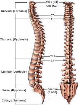
The vertebral column, also known as the spine or spinal column, is a series of bones called vertebrae that extend from the skull to the pelvis. It provides support for the body and protects the spinal cord. The five regions of the vertebral column, starting from the top and moving downwards, are:
- Cervical: This region is made up of seven vertebrae and is located in the neck. The first two cervical vertebrae, the atlas and the axis, are specialized to allow for head movement.
- Thoracic: This region is made up of twelve vertebrae and is located in the upper and middle back. The thoracic vertebrae are larger than the cervical vertebrae and articulate with the ribs.
- Lumbar: This region is made up of five vertebrae and is located in the lower back. The lumbar vertebrae are the largest and strongest of the vertebrae.
- Sacral: This region is made up of five fused vertebrae and is located in the pelvis. The sacrum forms the posterior wall of the pelvis and articulates with the hip bones.
- Coccygeal: This region is made up of four fused vertebrae and is located at the base of the vertebral column. The coccyx, or tailbone, provides atachment points for muscles and ligaments.
-
Q #4: What is the name of the genetic disorder caused by the presence of an extra chromosome 21?
A. Turner syndrome
B. Klinefelter syndrome
C. Down syndrome
D. Huntington's disease
Answer Explanation
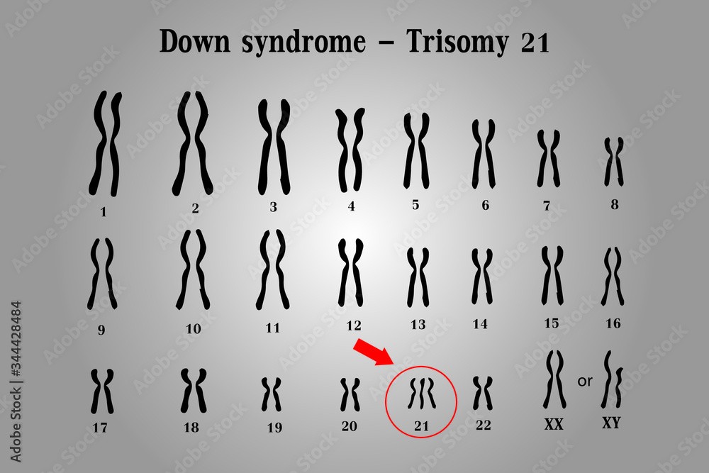
Down syndrome is a genetic disorder caused by the presence of an extra copy of chromosome 21. It is also known as trisomy 21, because affected individuals have three copies of chromosome 21 instead of the normal two.
The extra chromosome 21 in Down syndrome occurs due to a random error in cell division, which leads to the production of an abnormal gamete (egg or sperm) with an extra copy of the chromosome. When this gamete fuses with a normal gamete during fertilization, the resulting zygote has 47 chromosomes instead of the usual 46, and develops into a fetus with Down syndrome.
Down syndrome is characterized by a range of physical and intellectual symptoms, including developmental delays, intellectual disability, distinctive facial features, heart defects, and increased risk of certain medical conditions such as leukemia and Alzheimer's disease. However, the severity and expression of these symptoms can vary widely among affected individuals.
-
Q #5: What is the name of the valve that separates the left atrium and left ventricle in the heart?
A. Aortic valve
B. Mitral valve
C. Tricuspid valve
D. Pulmonary valve
Answer Explanation
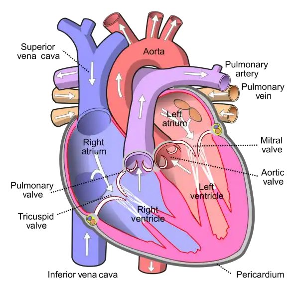
The mitral valve is located between the left atrium and left ventricle of the heart and helps to regulate the flow of blood between these chambers. It consists of two leaflets or flaps that open and close in response to changes in pressure as the heart beats.
During diastole, when the heart is relaxed and filling with blood, the mitral valve opens to allow blood to flow from the left atrium into the left ventricle. During systole, when the heart contracts to pump blood out of the left ventricle and into the systemic circulation, the mitral valve closes to prevent backflow of blood into the left atrium.
The mitral valve is one of four valves in the heart that help to ensure the unidirectional flow of blood through the heart and the rest of the circulatory system. Problems with the mitral valve, such as mitral valve prolapse or mitral stenosis, can lead to a range of symptoms and complications, including shortness of breath, fatigue, chest pain, and heart failure.
-
Q #6: What is the difference between isotonic and isometric muscle contractions?
A. Isotonic contractions produce no movement while isometric contractions produce movement.
B. Isotonic contractions produce movement while isometric contractions produce no movement.
C. Isotonic contractions generate tension in the muscle while isometric contractions involve shortening of the muscle fibers.
D. Isotonic contractions involve contraction of individual muscle fibers while isometric contractions involve the entire muscle.
Answer Explanation
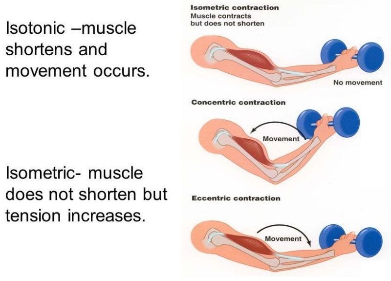
Isotonic and isometric contractions are two types of muscle contractions that differ in the amount of force produced and the movement of the muscle. In isotonic contractions, the muscle changes length and produces movement, such as lifting a weight. The force generated by the muscle remains constant throughout the movement. Isotonic contractions can be further classified as concentric contractions, in which the muscle shortens as it contracts, and eccentric contractions, in which the muscle lengthens as it contracts.
In contrast, isometric contractions occur when the muscle generates force without changing its length or producing movement. For example, holding a weight in a fixed position without moving it requires an isometric contraction. In an isometric contraction, the force generated by the muscle increases up to a maximum and then remains constant. Isometric contractions can be used to build strength and endurance in the muscle, but they do not produce movement.
-
Q #7: What is the difference between innate immunity and adaptive immunity?
A. Innate immunity is present at birth and provides immediate, non-specific protection against pathogens while adaptive immunity is developed over time and provides specific protection against particular pathogens.
B. Innate immunity involves the recognition of specific pathogens while adaptive immunity involves the recognition of general paterns of pathogens.
C. Innate immunity involves the production of antibodies while adaptive immunity involves the activation of phagocytes.
D. Innate immunity is activated by the lymphatic system while adaptive immunity is activated by the circulatory system.
Answer Explanation
Innate immunity is the first line of defense against pathogens and is present at birth. It provides immediate, non-specific protection against a wide range of pathogens, including bacteria, viruses, and fungi. Innate immunity involves physical barriers, such as skin and mucous membranes, as well as cellular and molecular components, such as phagocytes and cytokines.
Adaptive immunity, on the other hand, is developed over time and provides specific protection against particular pathogens. It involves the recognition of antigens, which are specific components of pathogens, by immune cells called lymphocytes. The lymphocytes then produce antibodies that are specific to the antigens, allowing for a targeted response to the pathogen. This process takes time to develop, as the immune system needs to encounter the pathogen and mount a response.
Overall, innate immunity provides immediate, non-specific protection while adaptive immunity provides specific protection that is tailored to the particular pathogen. Both forms of immunity work together to protect the body against pathogens.
-
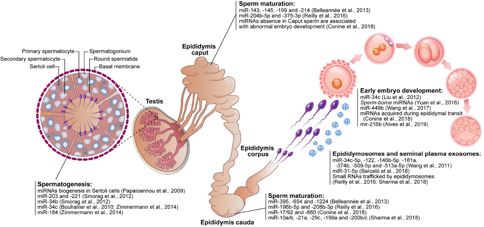
Q #8: What is the role of the epididymis in sperm maturation?
A. The epididymis produces sperm cells.
B. The epididymis stores and protects sperm cells until ejaculation.
C. The epididymis is responsible for the transport of sperm cells from the testes to the urethra.
D. The epididymis provides nourishment to sperm cells.
Answer Explanation
The epididymis is a coiled tube located at the back of each testicle where the sperm mature and are stored until ejaculation. Sperm are produced in the testes and then transported to the epididymis where they undergo maturation and become motile. The epididymis provides a protective environment for the sperm, allowing them to mature and become more resilient to external stressors. During ejaculation, the sperm are transported from the epididymis to the vas deferens and then to the urethra for ejaculation.
-
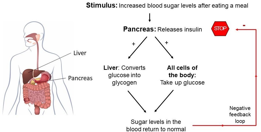
Q #9: What is the name of the hormone that regulates blood sugar levels in the human body?
A. Insulin
B. Glucagon
C. Estrogen
D. Testosterone
Answer Explanation
Insulin is a hormone produced by the pancreas that plays a crucial role in regulating the levels of glucose (sugar) in the blood. After a person eats a meal, the levels of glucose in the blood rise, which stimulates the pancreas to release insulin into the bloodstream. Insulin acts on various cells in the body, particularly those in the liver, muscles, and adipose tissue, to promote the uptake, use, and storage of glucose.
Insulin helps to lower the levels of glucose in the blood by increasing the uptake of glucose by cells, stimulating the liver and muscle cells to store glucose in the form of glycogen, and inhibiting the production and release of glucose by the liver. This process is known as glucose homeostasis, and it helps to keep the levels of glucose in the blood within a normal range.
Deficiencies or abnormalities in insulin production or function can lead to a range of metabolic disorders, including type 1 and type 2 diabetes. In type 1 diabetes, the body does not produce enough insulin, while in type 2 diabetes, the body becomes resistant to the effects of insulin, leading to elevated levels of glucose in the blood.
-
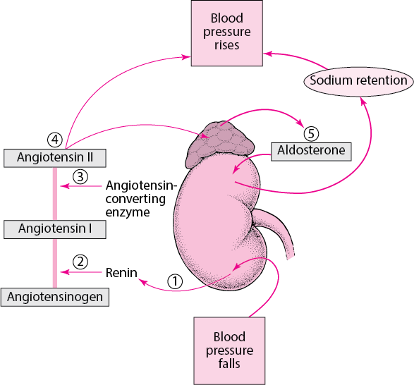
Q #10: Which of the following substances is excreted by the kidneys to regulate blood pressure?
A. renin
B. erythropoietin
C. calcitriol
D. urobilinogen
Answer Explanation
Renin is an enzyme that is produced by the kidneys and it acts to elevate blood pressure. When blood pressure falls, the kidneys secrete renin into the bloodstream ³.