Blood Flow
Blood Flow Through the Heart
The flow of blood through the heart is very orderly. It progresses through the heart to the lungs, where it receives oxygen; then goes back to the heart; and then out to the body tissues and parts.
The normal process of blood flow is:
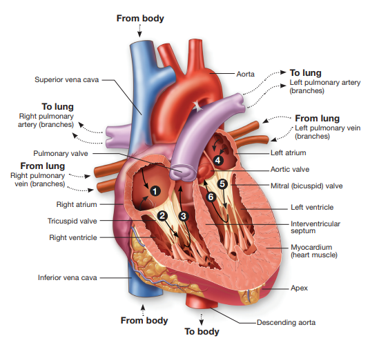
1. Deoxygenated blood from all the tissues in the body enters a relaxed right atrium via two large veins called the superior vena cava and inferior vena cava.
2. The right atrium contracts and blood flows through the tricuspid valve into the relaxed right ventricle.
3. The right ventricle then contracts and blood is pumped through the pulmonary valve into the pulmonary artery, which carries it to the lungs for oxygenation.
4. The left atrium receives blood returning to the heart after being oxygenated by the lungs. This blood enters the relaxed left atrium from the four pulmonary veins.
5. The left atrium contracts and blood flows through the mitral valve into the relaxed left ventricle.
6. When the left ventricle contracts, the blood is pumped through the aortic valve and into the aorta, the largest artery in the body. The aorta carries blood to all parts of the body. It can be seen that the heart chambers alternate between relaxing, in order to fill, and contracting to push blood forward. The period of time a chamber is relaxed is diastole. The contraction phase is systole.
Conduction System of the Heart
The heart rate is regulated by the autonomic nervous system; therefore, there is no voluntary control over the beating of the heart.
Special tissue within the heart is responsible for conducting an electrical impulse stimulating the different chambers to contract in the correct order. The path that the impulses travel is as follows:
1. The sinoatrial (SA, S-A) node, or pacemaker, is where the electrical impulses begin. From the sinoatrial node, a wave of electricity travels through the atria, causing them to contract, or go into systole.
2. The atrioventricular node is stimulated.
3. This node transfers the stimulation wave to the atrioventricular bundle (formerly called bundle of His).
4. The electrical signal next travels down the bundle branches within the interventricular septum.
5. The Purkinje fibers out in the ventricular myocardium are stimulated, resulting in ventricular systole.