Organs of The Alimentary Canal
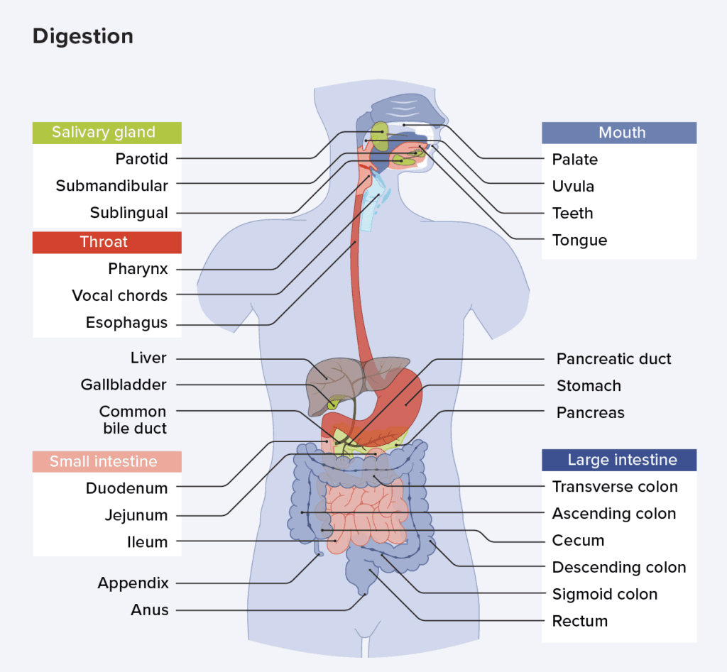
Mouth
Food enters the digestive tract through the mouth, or oral cavity, a mucous membrane-lined cavity.
-
Lips. The lips (labia) protect its anterior opening.
-
Cheeks. The cheeks form its lateral walls.
-
Palate. The hard palate forms its anterior roof, and the soft palate forms its posterior roof.
-
Uvula. The uvula is a fleshy finger-like projection of the soft palate, which extends inferiorly from the posterior edge of the soft palate.
-
Vestibule. The space between the lips and the cheeks externally and the teeth and gums internally is the vestibule.
-
Oral cavity proper. The area contained by the teeth is the oral cavity proper.
-
Tongue. The muscular tongue occupies the floor of the mouth and has several bony attachmentstwo of these are to the hyoid bone and the styloid processes of the skull.
-
Lingual frenulum. The lingual frenulum, a fold of mucous membrane, secures the tongue to the floor of the mouth and limits its posterior movements.
-
Palatine tonsils. At the posterior end of the oral cavity are paired masses of lymphatic tissue, the palatine tonsils.
-
Lingual tonsil. The lingual tonsils cover the base of the tongue just beyond.
Pharynx
From the mouth, food passes posteriorly into the oropharynx and laryngopharynx.
-
Oropharynx. The oropharynx is posterior to the oral cavity.
-
Laryngopharynx. The laryngopharynx is continuous with the esophagus below; both of which are common passageways for food, fluids, and air.
Esophagus
The esophagus or gullet, runs from the pharynx through the diaphragm to the stomach.
-
Size and function. About 25 cm (10 inches) long, it is essentially a passageway that conducts food by peristalsis to the stomach
-
Structure. The walls of the alimentary canal organs from the esophagus to the large intestine are made up of the same four basic tissue layers or tunics.
-
Mucosa. The mucosa is the innermost layer, a moist membrane that lines the cavity, or lumen, of the organ; it consists primarily of a surface epithelium, plus a small amount of connective tissue (lamina propria) and a scanty smooth muscle layer.
-
Submucosa. The submucosa is found just beneath the mucosa; it is a soft connective tissue layer containing blood vessels, nerve endings, lymph nodules, and lymphatic vessels.
-
Muscularis externa. The muscularis externa is a muscle layer typically made up of an inner circular layer and an outer longitudinal layer of smooth muscle cells.
-
Serosa. The serosa is the outermost layer of the wall that consists of a single layer of flat serous fluid-producing cells, the visceral peritoneum.
-
Intrinsic nerve plexuses. The alimentary canal wall contains two important intrinsic nerve plexuses- the submucosal nerve plexus and the myenteric nerve plexus, both of which are networks of nerve fibers that are actually part of the autonomic nervous system and help regulate the mobility and secretory activity of the GI tract organs.
Stomach
Different regions of the stomach have been named, and they include the following:
-
Location. The C-shaped stomach is on the left side of the abdominal cavity, nearly hidden by the liver and the diaphragm.
-
Function. The stomach acts as a temporary “storage tank” for food as well as a site for food breakdown.
-
Cardiac region. The cardiac region surrounds the cardioesophageal sphincter, through which food enters the stomach from the esophagus.
-
Body. The body is the midportion, and as it narrows inferiorly, it becomes the pyloric antrum, and then the funnel-shaped pylorus.
-
Pylorus. The pylorus is the terminal part of the stomach and it is continuous with the small intestine through the pyloric sphincter or valve.
-
Size. The stomach varies from 15 to 25 cm in length, but its diameter and volume depend on how much food it contains; when it is full, it can hold about 4 liters (1 gallon) of food, but when it is empty it collapses inward on itself.
-
Rugae. The mucosa of the stomach is thrown into large folds called rugae when it is empty.
-
Greater curvature. The convex lateral surface of the stomach is the greater curvature.
-
Lesser curvature. The concave medial surface is the lesser curvature.
-
Lesser omentum. The lesser omentum, a double layer of peritoneum, extends from the liver to the greater curvature.
-
Greater omentum. The greater omentum, another extension of the peritoneum, drapes downward and covers the abdominal organs like a lacy apron before attaching to the posterior body wall, and is riddled with fat, which helps to insulate, cushion, and protect the abdominal organs.
-
Stomach mucosa. The mucosa of the stomach is a simple columnar epithelium composed entirely of mucous cells that produce a protective layer of bicarbonate-rich alkaline mucus that clings to the stomach mucosa and protects the stomach wall from being damaged by acid and digested by enzymes.
-
Gastric glands. This otherwise smooth lining is dotted with millions of deep gastric pits, which lead into gastric glands that secrete the solution called gastric juice.
-
Intrinsic factor. Some stomach cells produce intrinsic factor, a substance needed for the absorption of vitamin b12 from the small intestine.
-
Chief cells. The chief cells produce protein-digesting enzymes, mostly pepsinogens.
-
Parietal cells. The parietal cells produce corrosive hydrochloric acid, which makes the stomach contents acidic and activates the enzymes.
-
Enteroendocrine cells. The enteroendocrine cells produce local hormones such as gastrin, that are important to the digestive activities of the stomach.
-
Chyme. After food has been processed, it resembles heavy cream and is called chyme.
Small Intestine
-
Location. The small intestine is a muscular tube extending from the pyloric sphincter to the large intestine.
-
Size. It is the longest section of the alimentary tube, with an average length of 2.5 to 7 m (8 to 20 feet) in a living person.
-
Subdivisions. The small intestine has three subdivisions: the duodenum, the jejunum, and the ileum, which contribute 5 percent, nearly 40 percent, and almost 60 percent of the small intestine, respectively.
-
Ileocecal valve. The ileum meets the large intestine at the ileocecal valve, which joins the large and small intestine.
-
Hepatopancreatic ampulla. The main pancreatic and bile ducts join at the duodenum to form the flasklike hepatopancreatic ampulla, literally, the ” liver-pancreatic-enlargement”.
-
Duodenal papilla. From there, the bile and pancreatic juice travel through the duodenal papilla and enter the duodenum together.
-
Microvilli. Microvilli are tiny projections of the plasma membrane of the mucosa cells that give the cell surface a fuzzy appearance, sometimes referred to as the brush border; the plasma membranes bear enzymes (brush border enzymes) that complete the digestion of proteins and carbohydrates in the small intestine.
-
Villi. Villi are fingerlike projections of the mucosa that give it a velvety appearance and feel, much like the soft nap of a towel.
-
Lacteal. Within each villus is a rich capillary bed and a modified lymphatic capillary called a lacteal.
-
Circular folds. Circular folds, also called plicae circulares, are deep folds of both mucosa and submucosa layers, and they do not disappear when food fills the small intestine.
-
Peyer’s patches. In contrast, local collections of lymphatic tissue found in the submucosa increase in number toward the end of the small intestine.
Large Intestine
The large intestine is much larger in diameter than the small intestine but shorter in length.
-
Size. About 1.5 m (5 feet) long, it extends from the ileocecal valve to the anus.
-
Functions. Its major functions are to dry out indigestible food residue by absorbing water and to eliminate these residues from the body as feces.
-
Subdivisions. It frames the small intestines on three sides and has the following subdivisions: cecum, appendix, colon, rectum, and anal canal.
-
Cecum. The saclike cecum is the first part of the large intestine.
-
Appendix. Hanging from the cecum is the wormlike appendix, a potential trouble spot because it is an ideal location for bacteria to accumulate and multiply.
-
Ascending colon. The ascending colon travels up the right side of the abdominal cavity and makes a turn, the right colic (or hepatic) flexure, to travel across the abdominal cavity.
-
Transverse colon. The ascending colon makes a turn and continuous to be the transverse colon as it travels across the abdominal cavity.
-
Descending colon. It then turns again at the left colic (or splenic) flexure, and continues down the left side as the descending colon.
-
Sigmoid colon. The intestine then enters the pelvis, where it becomes the S-shaped sigmoid colon.
-
Anal canal. The anal canal ends at the anus which opens to the exterior.
-
External anal sphincter. The anal canal has an external voluntary sphincter, the external anal sphincter, composed of skeletal muscle.
-
Internal involuntary sphincter. The internal involuntary sphincter is formed by smooth muscles.