The Heart
The heart, a muscular pump made up of cardiac muscle fibers, could be considered a muscle rather than an organ. It has four chambers, or cavities, and beats an average of 60–100 beats per minute (bpm) or about 100,000 times in one day.
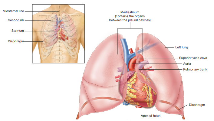
Each time the cardiac muscle contracts, blood is ejected from the heart and pushed throughout the body within the blood vessels. The heart is located in the mediastinum in the center of the chest cavity; however, it is not exactly centered; more of the heart is on the left side of the mediastinum than the right.
At about the size of a fist and shaped like an upside-down pear, the heart lies directly behind the sternum. The tip of the heart at the lower edge is called the apex.
Layers of the Heart
The wall of the heart is quite thick and is composed of three layers:
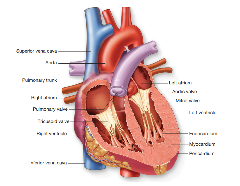
1. The endocardium is the inner layer of the heart lining the heart chambers. It is a very smooth, thin layer that serves to reduce friction as the blood passes through the heart chambers.
2. The myocardium is the thick, muscular middle layer of the heart. Contraction of this muscle layer develops the pressure required to pump blood through the blood vessels.
3. The epicardium is the outer layer of the heart. The heart is enclosed within a double-layered pleural sac, called the pericardium.
The epicardium is the visceral pericardium, or inner layer of the sac. The outer layer of the sac is the parietal pericardium. Fluid between the two layers of the sac reduces friction as the heart beats.
Hear Chambers
The heart is divided into four chambers or cavities.
There are two atria, or upper chambers, and two ventricles, or lower chambers.
These chambers are divided into right and left sides by walls called the interatrial septum and the interventricular septum. The atria are the receiving chambers of the heart. Blood returning to the heart via veins first collects in the atria.
The ventricles are the pumping chambers. They have a much thicker myocardium and their contraction ejects blood out of the heart and into the great arteries.
Heart Valves
Four valves act as restraining gates to control the direction of blood flow. They are situated at the entrances and exits to the ventricles.
Properly functioning valves allow blood to flow only in a forward direction by blocking it from returning to the previous chamber.
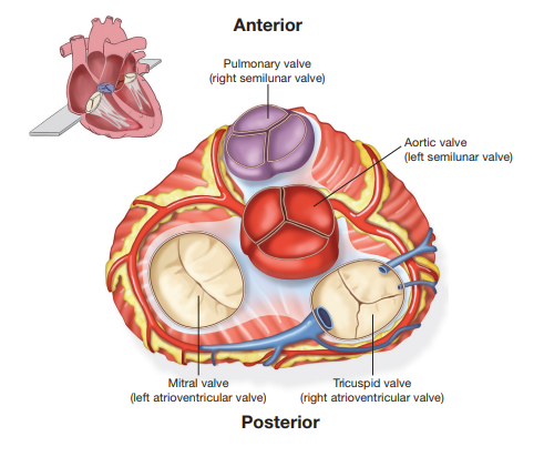
The four valves are:
1. Tricuspid valve: an atrioventricular valve (AV), meaning that it controls the opening between the right atrium and the right ventricle. Once the blood enters the right ventricle, it cannot go back up into the atrium again. The prefix tri-, meaning three, indicates that this valve has three leaflets or cusps.
2. Pulmonary valve: a semilunar valve, with the prefix semi- meaning half and the term lunar meaning moon, indicate that this valve looks like a half moon. Located between the right ventricle and the pulmonary artery, this valve prevents blood that has been ejected into the pulmonary artery from returning to the right ventricle as it relaxes.
3. Mitral valve: also called the bicuspid valve, indicating that it has two cusps. Blood flows through this atrioventricular valve to the left ventricle and cannot go back up into the left atrium.
4. Aortic valve: a semilunar valve located between the left ventricle and the aorta. Blood leaves the left ventricle through this valve and cannot return to the left ventricle.